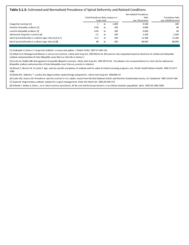
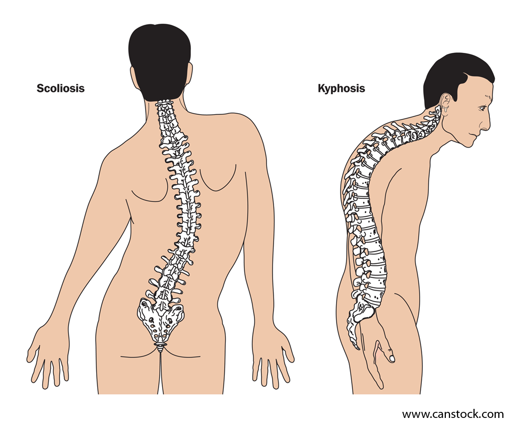
Infantile scoliosis currently accounts for less than 1% of all cases of idiopathic scoliosis in the United States. Boys are affected by infantile idiopathic scoliosis at a higher rate than girls (3:2 ratio).1 Infantile scoliosis curves tend to be left-sided (75% to 90%). Past studies have indicated this rare type of scoliosis occurs more frequently in Europe than in North America.2
Treatment for patients with infantile idiopathic scoliosis is determined by anticipated or actual curve progression. Several common measurement techniques are used, with angles ≤20° generally considered at low risk for progression. Re-evaluation is recommended every 4 to 6 months.1
In addition to measuring the Cobb angle, the RVAD is used as a common predictor of curve progression.3 Patients with a Cobb angle of ≤25° and a RVAD of ≤20° are at a low risk for progression and should be re-evaluated every 4 to 6 months.1
Nonsurgical treatment, such as bracing or casting is initiated if a curve progression of ≥10° occurs. Surgical treatment should be considered when nonsurgical measures, including both bracing and casting, are not successful. Surgical treatment is utilized when a curve is ≥45° and progressive in an immature child.1 Overall, surgical methods are continually evolving, with the goal of obtaining and maintaining curve correction while simultaneously preserving or encouraging spinal and trunk growth.
Surgical options currently utilized include various types of spinal fusion or hemiepiphysiodesis, a minimally invasive implant procedure to slow progression of curve growth. Additional techniques include growing-rod instrumentation (rods that expand and support the deformed spine) and vertical expandable (telescoping) prosthetic titanium rib (VEPTR) instrumentation.4The goal of using surgical methods is to halt the progression of the curve and gain correction of the deformity, allowing maximum growth of the spine, lungs, and thoracic cage.1
- 1. a. b. c. d. e. Akbarnia B: Management themes in early onset scoliosis. J Bone Joint Surg Am 2007;89:42-54.
- 2. Fernandes P, Weinstein SL: Natural history of early onset scoliosis. J Bone Joint Surg Am 2007;89:21-33.
- 3. Mehta M: The rib-vertebra angle in the early diagnosis between resolving and progressive infantile scoliosis. J Bone Joint Surg Br 1972;54:230-243.
- 4. Titanium Rib or VEPTR (Vertical Expandable Prosthetic Titanium Rib): An expandable titanium metal rod placed in a vertical position alongside the spine and attached to the ribs and pelvis or the spine. The VEPTR expands and supports a deformed chest wall cavity giving the lungs room to operate and grow. VEPTR is a new method for the treatment of thoracic insufficiency syndrome (TIS) and congenital spinal deformity (scoliosis) in children. Available at: http://www.ncbi.nlm.nih.gov/pubmed/15931035 . Accessed October 22, 2014.
Edition:
- 2014


 Download as CSV